| Technical Name | Intelligent recognition display for endoscope | ||
|---|---|---|---|
| Project Operator | National Chung Cheng University | ||
| Project Host | 王祥辰 | ||
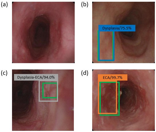
| Summary | Esophagogastroduodenoscopy is the most sensitive examination method for esophageal cancer diagnosis, but the diagnosis of precancerous lesions (high-grade dysplasia and carcinoma in situ) and superficial esophageal cancer still present great challenge to endoscopists because these lesions are easily ignored in conventional white-light imaging (WLI). Even though image-enhanced endoscopies (IEEs), including dye-based and equipment-based endoscopies, are usually recommended in addition to WLI in improving the detection rate of esophageal superficial neoplasm. The SSD model took 10 seconds to analyze 264 test images and accurately detected the 224 images of esophageal symptoms, and the SSD diagnosis accuracy of the WLI images was 83%. Sensitivity according to the severity of esophageal cancer had the following values: 78% (normal), 61% (dysplasia), 86% (dysplasia-ECA), and 93% (ECA), with accuracies of 72%, 85%, 86%, and 85%, respectively. |
||
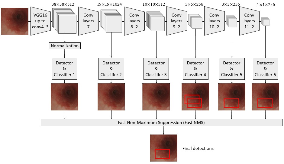
| Scientific Breakthrough | In the context of medical imaging, deep learning can become a powerful support tool that can interpret medical images based on a cumulative set of unique algorithms. Deep learning allows a computing model composed of multiple processing layers to learn data representations with multiple levels of abstraction. Here, we provide a set of single-step multi-frame detection based on Convolutional neural network (CNN) The Single Shot Multibox Detector (SSD) for esophageal cancer image recognition system proves that AI has the diagnostic ability to detect esophageal cancer, helps doctors identify the symptoms of esophageal cancer under WLI and NBI, and reduces the risk of subjective misjudgment. |
||
| Industrial Applicability | It is used in the relevant medical institutions and clinics, and is equipped with an endoscopy diagnosis system to quickly and accurately diagnose the patient's disease occurrence area and severity. |
||
| Keyword | esophageal cancer single-shot multibox detector artificial intelligence convolutional neural network | ||
- hcwang@ccu.edu.tw
other people also saw


